Monitoring Ventilator-Associated Events: Facilitator Guide
AHRQ Safety Program for Mechanically Ventilated Patients
Slide 1: Monitoring Ventilator-Associated Events
Say:
Today, we will discuss monitoring ventilator-associated events.
Slide 2: Learning Objectives
Say:
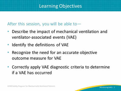
After this session, you will be able to describe the impact of mechanical ventilation and ventilator-associated events or VAEs, identify the definitions of the three VAE tiers, recognize the importance of monitoring VAE and objective outcome measures, and correctly apply VAE diagnostic criteria to determine if a VAE has occurred.
Slide 3: Impact of Mechanical Ventilation
Ask:
Why focus this improvement program on mechanically ventilated patients?
Say:
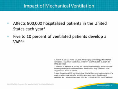
Approximately 800,000 hospitalized patients are mechanically ventilated in the United States each year and of these, 5 to 10 percent—between 40,000 and 80,000—develop some type of VAE.
Slide 4: Impact of Mechanical Ventilation

Say:
Historically, ventilator-associated pneumonia, or VAP, was considered one of the most lethal healthcare-associated infections. On average, there is a 35 percent mortality rate for ventilated patients—24 percent for patients 15-19 years old but rising to 60 percent for patients 85 years old or older. VAP contributes to this mortality risk.
Slide 5: Possible Complications of Mechanical Ventilation

Say:
Mechanical ventilation puts patients at risk for a number of complications, including ventilator-associated events, acute respiratory distress syndrome, sepsis, pulmonary embolism and edema, barotrauma, and more. There is some overlap of categories, as some of these complications cause oxygenation changes that qualify for ventilator-associated events, or VAEs. VAEs can be categorized into three tiers, which we will address later in this module. Those categories are ventilator-associated conditions or VAC, infection-related ventilator-associated conditions or IVAC, and ventilator-associated pneumonia or VAP.
Slide 6: Understanding the Impact of VAC
Say:
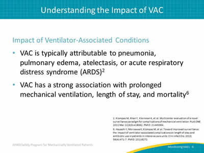
VAC is the first category of VAE. Five to 10 percent of ventilated patients suffer from some type of VAC, typically attributable to pneumonia, pulmonary edema, atelectasis, or acute respiratory distress syndrome, ARDS. Multiple studies have shown a strong association between VACs and prolonged mechanical ventilation, length of stay, and mortality.
Slide 7: Understanding the Impact of VAC
Say:
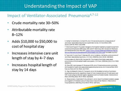
The classic quality measure for mechanically ventilated populations is VAP. This is because VAP affects a large number of patients. It increases length of stay in the intensive care unit and the hospital by a substantial amount, has a very high crude mortality rate and a high attributable mortality rate, and adds substantially to cost.
Slide 8: Why Is This Work Important?

Ask:
First, let’s ask ourselves, "Why is this work important?"
Say:
Not only do VAEs contribute considerably to an increase in days to extubation and days to hospital discharge, but suffering a VAE doubles the patient’s risk of dying compared to a similarly vented patient who did not experience a VAE.
Slide 9: National Health Safety Network VAE Definition

Say:
In 2013, the Centers for Disease Control and Prevention developed the National Healthcare Safety Network, or NHSN, VAE Surveillance Definition. Prior to 2013, such surveillance focused solely on VAP and definitions were considered to be subjective and difficult to use. In addition, focusing solely on VAP prevented a valid and reliable means of surveilling the data that is considered necessary for assessing the effectiveness of prevention strategies on the outcomes of patients on mechanical ventilation not specifically suffering from VAP. Thus, the new NHSN VAE Surveillance Definition was created.
The new definitions, broken down into tiers, are objective, streamlined, potentially automatable with algorithms developed for Epic or other electronic medical records, and define a broad range of conditions and complications occurring in mechanically ventilated patients.
Slide 10: What Types of Mechanical Ventilation Are Included?

Say:
All types of mechanical ventilation are included in the NHSN VAE definition, with the exception of—
- High-frequency ventilation.
- Extracorporeal membrane oxygenation.
- Non-invasive lung expansion devices, such as—
- Intermittent positive pressure breathing.
- Nasal positive end-expiratory pressure (PEEP).
- Nasal continuous positive airway pressure.
- Airway pressure release ventilation (APRV) and related modes are included, however, only fraction of inspired oxygen (FiO2) values are used.
Slide 11: VAE Definition Tiers
Say:
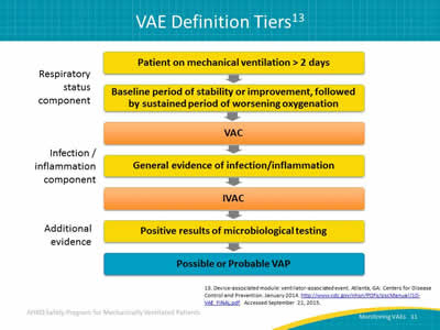
Here you see the VAE definitions tiers. At the top is the respiratory status component. In this tier, you will see a baseline period of stability or improvement in oxygenation then followed by a sustained period of worsening oxygenation. This is the definition for VAC. The next tier is the infection/inflammation component. Here, you will see generalized evidence of infection or inflammation which is considered the definition for IVAC. Finally, with the benefit of additional evidence, such as the positive results of microbiological testing, you can determine if there is possible VAP.
Slide 12: VAC—Definition Criteria
Say:
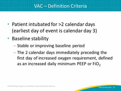
In order to determine VAC, the patient must meet the following conditions. They must have been intubated for at least 2 days prior to determination. The earliest a patient can experience an event is the third calendar day of intubation. Additionally, the patient must have a period of baseline stability. The 2 calendar days immediately preceding the day the patient experiences worsening oxygenation (defined as an increased daily minimum PEEP or FiO2) must show the patient stable or improving.
Slide 13: VAC—Determination

Ask:
What is considered "worsening oxygenation"?
Say:
The indicators of worsening oxygenation for the purposes of determining VAC are—
- Daily minimum PEEP values increase ≥ 3 cm H2O over daily minimum for the preceding 2 calendar days.
- Daily minimum FiO2 values increase ≥ 0.20 over daily minimum for preceding 2 calendar days.
- PEEP or FiO2 must be maintained for ≥ 1 hour (two consecutive hour readings).
Slide 14: VAC—Exceptions to One-Hour Rule

Ask:
What if our unit is unable to record hourly PEEP or FiO2 values?
Say:
There are exceptions to the "1-hour" rule in determining worsening oxygenation. If the PEEP or FiO2 values are not recorded hourly, you should use the lowest value. If the PEEP or FiO2 values are not stable for at least an hour (such as when a patient is extubated early in the day or admitted late in the day), use the lowest value.
Slide 15: IVAC—Criterion 1
Say:
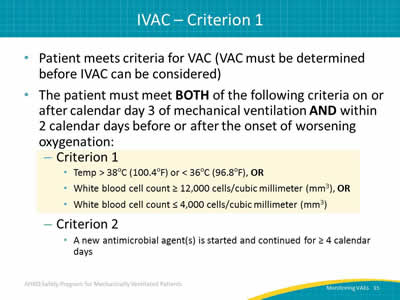
In order to meet criteria for IVAC, the patient must first meet criteria for VAC. Once VAC has been established using the criteria we just reviewed, the patient can be evaluated for IVAC. Additionally, the patient must meet the two IVAC criterions on or after calendar day three of mechanical ventilation and within 2 days before or after the onset of worsening oxygenation.
The first criterion is that the patient must have a temperature greater than 38 degrees Celsius (100.4 degrees Fahrenheit) or less than 36 degrees Celsius (96.8 degrees Fahrenheit). Or, the patient must have a white blood cell count greater than or equal to 12,000 cells per cubic millimeter or less than or equal to 4,000 cells per cubic millimeter.
Slide 16: IVAC—Criterion 2

Say:
The second criterion for IVAC is that a new antimicrobial agent must have been started and continued for at least four calendar days. We will discuss what constitutes a new microbial agent over the next few slides.
Slide 17: IVAC Antimicrobial Criteria

Say:
Antimicrobial criteria for determining IVAC can be complicated; however, these criteria standardize the assessment method of antimicrobial therapy even with some information unavailable. This means IVAC can be determined even if the status of renal function, as well as antimicrobial dosing and indication, are all unknown.
Slide 18: IVAC—Antimicrobials Included

Say:
In the past, there was a broad range of agents for all healthcare-acquired infections—not just respiratory infections—included in this criteria. Currently, the antimicrobials included in the criteria are antibacterials, antifungals, and limited antivirals.
Slide 19: IVAC—Antimicrobials NOT Included
Say:
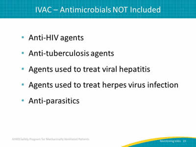
Antimicrobials that cannot be used to diagnose IVAC are anti-HIV agents, anti-tuberculosis agents, agents used to treat viral hepatitis, agents used to treat herpes virus infections, and anti-parasitics.
Slide 20: Definition—New Antimicrobial Agent
Say:
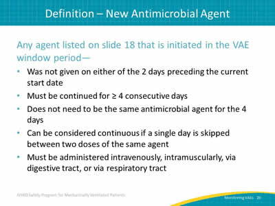
A new antimicrobial agent is any agent (with the exception of those listed on the following slide) initiated in the VAE window period. It cannot be administered on either of the 2 days preceding the current start date of the VAE. The agent must be continued for at least 4 consecutive days. Each of these consecutive days is referred to as a "qualifying antimicrobial day" or QAD. However, it does not need to be the same antimicrobial agent for 4 days, provided the patient is on at least one new antimicrobial agent on each QAD. Days can still be considered continuous if only a single day is skipped ONLY between two doses of the same agent. Additionally, it must not have been given on either of the 2 days preceding the current start date, and must be administered intravenously, intramuscularly, orally, or through the respiratory tract.
Slide 21: IVAC—New Antimicrobials NOT Included
Say:
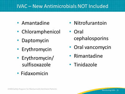
The antimicrobial agents listed here should not be included in the assessment of IVAC since they are not likely to be used to treat a lower respiratory infection in critically ill patients.
Slide 22:

Say:
We will now discuss the importance of objective outcomes when diagnosing VAP and monitoring VAEs.
Slide 23: Diagnostic Criteria for VAP
Say:
As we start to think about our capacity to use VAP as a quality metric and our capacity to track whether better care leads to better outcomes with patients, there are number of problems.
Ask:
So, the first question you might ask is, "How should you define ventilator-associated pneumonia?"
Say:
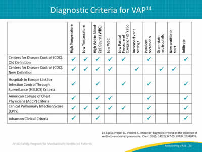
A study published in the journal Chest in 2014 found six definitions for ventilator-associated pneumonia; they are laid out for us on this slide. You can see that all these definitions are different. They all have different components to them, and therefore, it should not surprise you to see that they all lead to very different apparent VAP rates, as shown on the next slide.
Slide 24: Impact of Diagnostic Criteria on VAP Prevalence

Say:
Here you can see different rates according to which definition you might choose to use. Unfortunately, it is unknown if one of these definitions is any better than another, and we have no information to tell us if one of these definitions is more accurate than another.
Ask:
Why is this a problem?
Say:
The problem is that all the signs used to define VAP are very subjective because they are not specific to VAP. The core clinical signs associated with VAP used for the diagnosis are radiographic opacities, fever, abnormalities of the white blood cell count, impaired oxygenation, and increased secretions. If you think about each of those signs, you will see a lot of subjectivity with a broad differential diagnosis.
Slide 25: Inconsistency in VAP Diagnoses

Say:
If you are trying to use VAP as your outcome measure to see if your quality improvement initiative is working or not, you may encounter some of these problems. Even clinicians have a lot of disagreement at the bedside over who does and does not have a pneumonia.
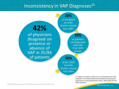
The figure you see comes from a study out of France that examined a series of 84 patients with abnormal chest x rays and purulent sputum. Often, this combination would indicate a pneumonia, and an initial reaction would be to prescribe broad spectrum antibiotics. In this study, they had those patients evaluated independently by seven different doctors and they tried to establish a true diagnosis either by histology or quantitative bronchoscopy cultures. The first takeaway from this research was that despite those tools for diagnosis, only a minority of patients, 32 percent, who appeared to have VAP actually had VAP.
Slide 26: Inconsistency in VAP Diagnoses
Say:
The second takeaway is that there was a great deal of disagreement. The physicians disagreed on the presence or absence of VAP in 42 percent of the patients. Furthermore, it was not simply that some doctors agreed a lot with others and some other doctors did not agree much. The very "best" doctors still missed a quarter of the true VAPs. The "worst" doctors missed half of the true VAPs. And, both groups mislabeled about 20 percent of patients without a VAP as having a VAP. We can see that the error goes in both directions, both undercalling and overcalling.
The bottom line is that the old clinical diagnosis for VAP is simply poor. As much as we think we might be able to tell who does and does not have a pneumonia, the evaluation studies do not support that belief.
Slide 27: AHRQ Safety Program for Mechanically Ventilated Patients

Say:
We will now use the VAC criteria reviewed earlier to determine whether a VAC has occurred.
Slide 28: Let’s Find a VAC

Say:
Here is a refresher on the clinical criteria used to determine whether a patient is experiencing a VAC.
Ask:
Do you remember the definition of "worsening oxygenation"?
Say:
Worsening oxygenation is indicated when—
- Daily minimum PEEP values increase ≥ 3 cm H2O over daily minimum for the preceding 2 calendar days.
- Daily minimum FiO2 values increase ≥ 0.20 over daily minimum for preceding 2 calendar days.
Slide 29: Example 1__PEEP

Ask:
In this first example, the patient’s minimum PEEP was five on ventilation days five and six. On day seven, it increased to eight, and on day nine, it increased to nine.
Say:
Is this a VAC?
Slide 30: Example 1__PEEP

Say:
Yes! This is a VAC. As you can see, the patient had a stable PEEP for days five and six. On day seven, the PEEP increased to eight and remained there for 2 days. Because the PEEP increased by three and remained there for 2 days, this is considered a VAC.
Slide 31: Example 2__FiO2
Say:
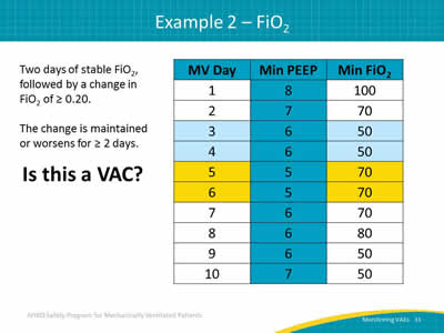
In our second example, the patient’s minimum FiO2 was fifty on days three and four. On day five, it increased to 70 and remained there on day six.
Ask:
Is this a VAC?
Slide 32: Example 2__FiO2
Say:
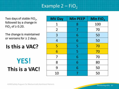
Yes! This is a VAC. The patient had a stable FiO2 on days three and four. On day five, the FiO2 increased to 70 and remained high for the next 3 days. Because the FiO2 increased by greater than 20 percent and remained there for more than 2 days, this is considered a VAC.
Slide 33: Example 3

Say:
Here is our last example. For this one, let’s look at both the PEEP and FiO2 values at the same time. Take a moment to look over these measurements and decide whether this example is a VAC.
Ask:
Is this a VAC?
Slide 34: Example 3
Say:
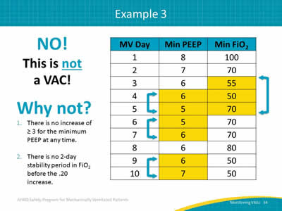
No! This is NOT a VAC.
Ask:
Why not?
Say:
There is no increase of three or greater for the minimum PEEP value at any time; the three changes recorded only increases by one. There is no 2-day stability period in FiO2 before the 20 percent increase between days four and five. Since neither of the VAC criteria have been met, this patient has not experienced a VAC.
Slide 35: Questions?

Say:
Does anyone have any questions about the material we have just reviewed?
Slide 36: References
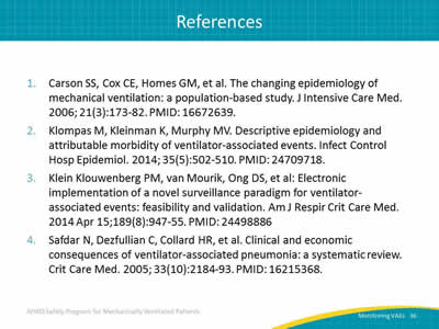
Slide 37: References

Slide 38: References
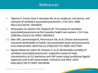
Slide 39: References


